Selected Research
Here are a couple of past projects. For a complete list, please see my publications and CV or resume.Segmentation despite extreme anatomical variability
At MIT, with Polina Golland, Mehdi Hedjazi Moghari, Adrian Dalca, Jürgen Weese, Tom Brosch, Tal Geva and Andrew Powell.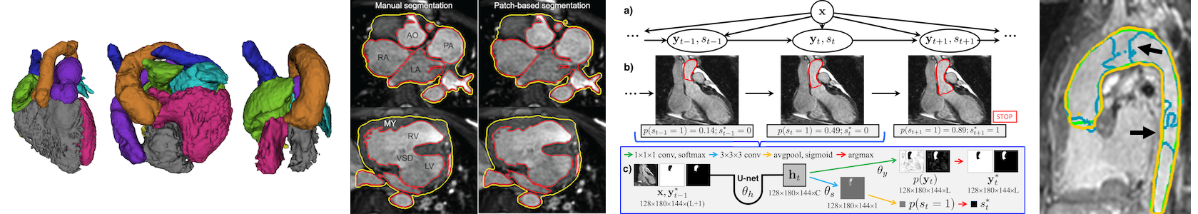
My Ph.D. work was in machine learning for image segmentation, specifically for challenging clinical applications involving wide anatomical variability and little training data. My main focus was on segmenting the cardiac chambers and great vessels in 3D MRI scans from children with congenital heart disease (CHD). CHD encompasses a broad range of heart defects, and involves significant morphological and topological changes to the anatomy of the heart.
To address this, I developed new segmentation methods involving interactive segmentation, probabilistic modeling and machine learning. For example, our most recent work develops a model for iterative segmentation which evolves a segmentation over several steps, and implements it as a recurrent neural network, improving generalization to patients with severe cardiac malformations.
Sliding organ registration
At Kitware, with Stephen Aylward and Marc Niethammer.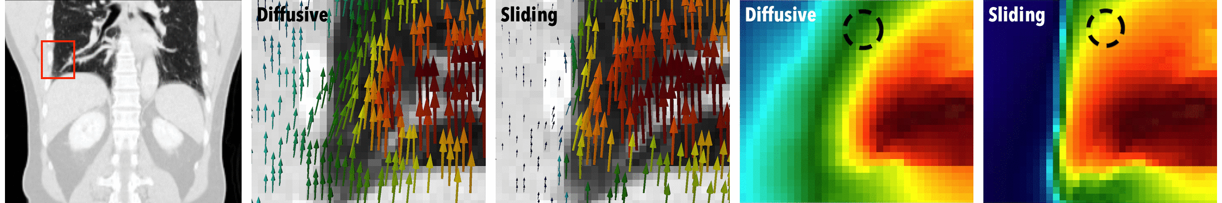
Conventional image registration techniques encourage smooth transformations, but this prior is not appropriate when organs move independently. For example, the lungs slide along the chest wall during breathing: look closely, and you can see that the lungs and abdominal organs move while the ribs are stationary. We proposed a deformable image registration algorithm that uses anisotropic smoothing for regularization. Our method recovers the expected motion discontinuities caused by sliding, while also improving the alignment of the lungs during respiratory motion compensation.
4D ultrasound reconstruction for computer assisted interventions
At the University of Western Ontario, advised by Terry Peters.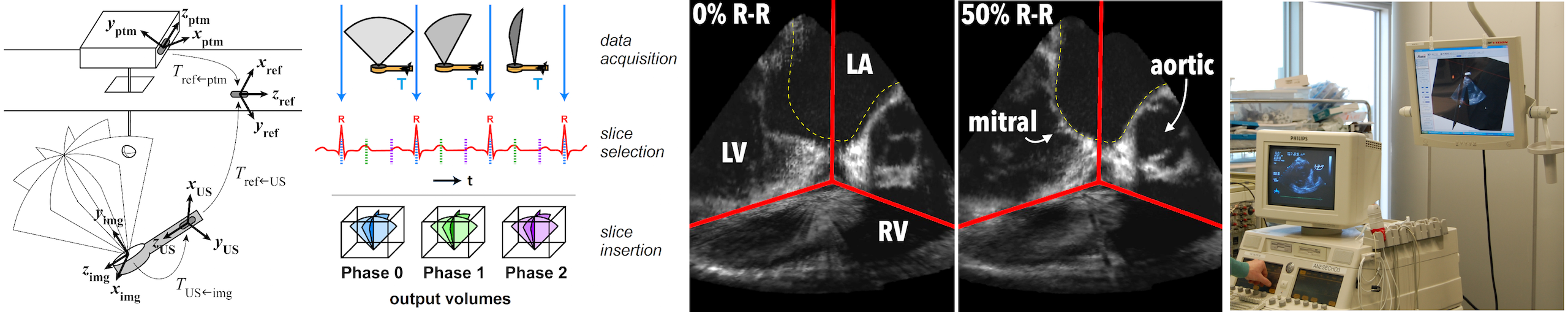
Intraoperative imaging is essential during minimally-invasive cardiac interventions, since direct visualization of the heart is impossible. We developed a gated 4D ultrasound reconstruction approach and incorporated it within an augmented reality environment for surgical guidance. A tracked 2D ultrasound probe is used to generate a time series of 3D images representing the beating heart, which can be used for visualization or for registration to preoperative data. Validation was performed in the laboratory and all the way to evaluation in actual cardiac patients.
https://som.ucdenver.edu/Profiles/Faculty/Profile/35789