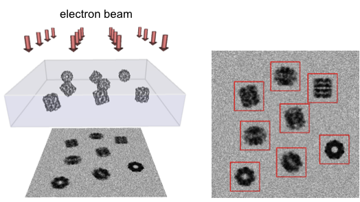
Cryo-EM imaging (left) and boxing (right)
Cryo-electron microscopy (cryo-EM)
Cryo-EM is a method that uncovers the structure of macromolecular complexes. It involves purifying a solution of a certain macromolecular complex, placing the solution onto a grid, freezing it (cryogenically), and then imaging the frozen film using an electron microscope. This produces many images of the same complex in different orientations, as shown in the image below.
Cryo-EM imaging (left) and boxing (right)
In single-particle reconstruction, the image of each complex is first separated, by boxing, or specifying a rectangle that tightly bounds it in the larger image. All the boxed images are then clustered. The images in each cluster are averaged, to improve the signal-to-noise ratio. The image below shows three clusters, with images from each cluster and the averaged image on the same row. The 3D orientation of each of these average images is then found. Using these orientations, the images are back-projected to produce a 3D density map. This 3D density map captures the electron density throughout the macromolecular complex.
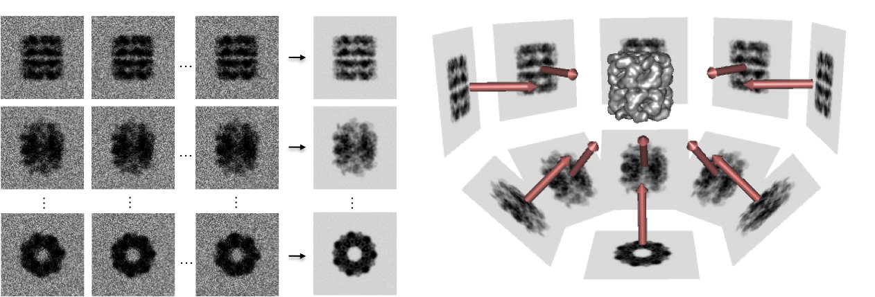
Cryo-EM clustering (left) and back-projection (right)
References