| D. Gering. Flattened Anatomy for Interactive Segmentation and Measurement. SIGGRAPH Poster, San Diego, CA, July 2007. |
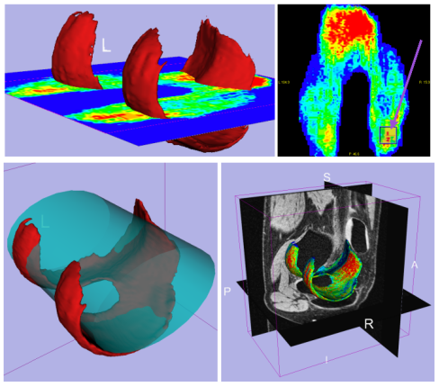
| ABSTRACT: Many anatomical surfaces can be described as 2-D manifolds embedded within 3-D space. The manifolds could be linear, such as a plane, or non-linear, such as a curved sheet. In some applications, it would be desirable for the user to be able to interact with the manifold by drawing upon it in some way. The purpose of this drawing could be to perform quantitative measurements such as to measure distances, surface areas, or volumes. The purpose could also be to analyze local properties of the 3-D surface at specific locations, where these properties include thickness and curvature. While methods exist for flattening certain anatomies for simple viewing, we propose enabling the user to interact with the flattened manifold. Furthermore, we augment the flattened surface to be a map of surface properties, such as thickness, curvature, or abnormality. |
 |
| 406K |
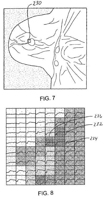
| D. Gering, J. Hoford. Graphlets: A Method for Visualizing Dynamic Data. SIGGRAPH Poster, San Diego, CA, July 2007. |
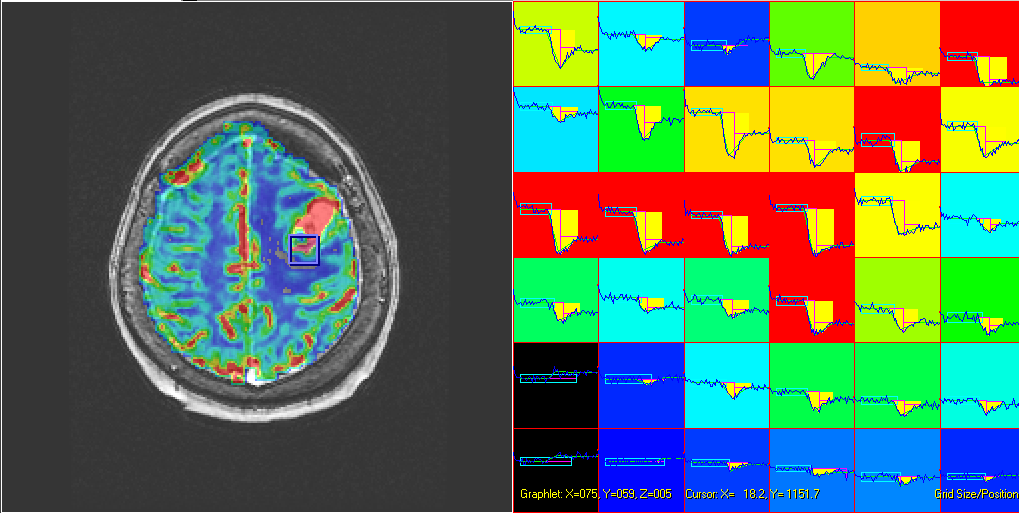
| ABSTRACT: A new method for augmented visualization of dynamic data is presented. An acquired MRI slice is displayed side-by-side with a 2-D array of miniature graphs, referred to as graphlets. Each graphlet plots a curve that graphs signal intensity over time for a corresponding spatial location. The color of each graphlet's backdrop is determined from either the original MRI signal, or the computed parametric result. The user can conveniently navigate -- selecting which region of the slice is rendered as graphlets -- by using the mouse for real-time zooming and panning. |
|
| 172K |
| D. Gering. A Flight Simulator Approach to the Visualization of Dynamic Medical Data. SIGGRAPH Poster, Boston, MA, July 2006. |
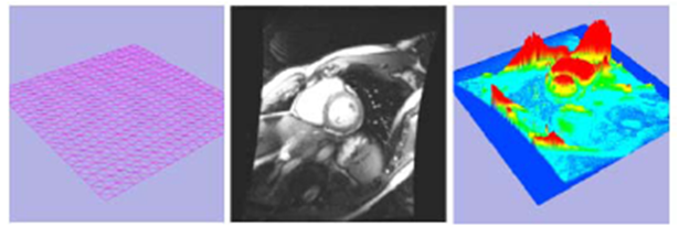
| ABSTRACT: A method was presented for visualizing medical data by virtually flying through it as if the data represented dynamic terrain in a flight simulator. Contrast between data points is made apparent with respect to both color and elevation, thereby giving the user a different perspective than what can be shown using color alone. |
|
| 120K |
| D. Gering. Interactive Animations for Medical Data Visualization. SIGGRAPH Poster, Boston, MA, July 2006. |
| ABSTRACT: A method was presented that makes it convenient for a physician to record a script that can be used for offline rendering, and an interface for combining real-time interaction with a stored animation. Users can have both real-time interaction (to determine which navigation motions best explore the data for the case at hand), and the highest possible image quality for the stored animation (the level that can only be produced by a render farm). This combination offers a reviewing physician the choice of simply watching the high-quality animation unfold, or interrupting it to temporarily navigate the 3-D scene before continuing with the animation in the most seamless manner. |
|
| 105K |
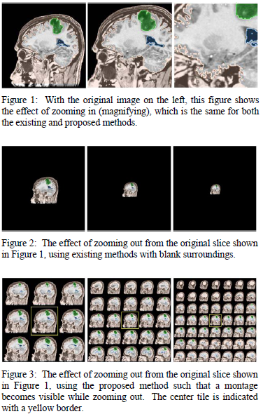
| D. Gering. Integrated Image Zoom and Montage. SIGGRAPH Poster, Boston, MA, July 2006. |
| ABSTRACT: A method was presented that allows the user to rapidly transition between bird�s-eye views and close-up views, which could aid the user�s understanding of the dataset. The user zooms out into a montage of proximal slices instead of a blank surrounding. From here, the user may pan in order to zoom in on a new slice. |
|
| 196K |
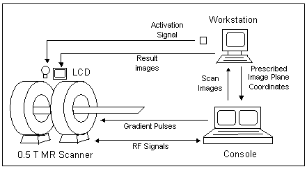
| D. Gering, D. Weber. Intraoperative, Real-time, Functional MRI. Journal of Magnetic Resonance Imaging (JMRI), 8(1):254-257, January 1998. |
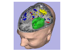
| ABSTRACT: Functional MRI (fMRI) methods have been demonstrated to noninvasively identify motor-sensory, visual, and other areas of eloquent cortex for guiding surgical intervention. Typically, fMRI data are acquired preoperatively during a conventional surgical planning MRI examination. Unlike direct cortical stimulation at the time of surgery, however, preoperative fMRI methods do not account for the potential movement of tissues (relative to the time of functional imaging) that may occur in the surgical suite as a direct result of the intervention. Recently, an MRI device has been demonstrated for use in the surgical suite that has the potential to reduce the extent of cortical exposure required for the intervention. However, the invasive requirements of cortical mapping may supersede the invasive requirements of the surgical intervention itself. Consequently, we demonstrate here a modification to the intraoperative MRI device that facilitates a noninvasive, real-time, functional MR examination in the surgical suite. |
|
| 1.2M |
| D. Gering. Systems and Methods for Interactive Image Registration. US Patent #US7693349, General Electric Co., August 2006. |
| ABSTRACT: System and method for incorporating user input on the fly during an otherwise automatic registration process. During rigid registration, user input adjusts the current computed pose or transformation that relates the two images being aligned. During warping, user input adjusts the flow field locally, and is gradually smoothed into the surrounding flow field. During multi-scale registration where images are first aligned at a course resolution, and subsequently at progressively finer resolutions, user input is applied at the current scale. User input is detected during the automated process either by interrupts or polling. Between user inputs the registration results are re-rendered. |
|
| 2.2M |
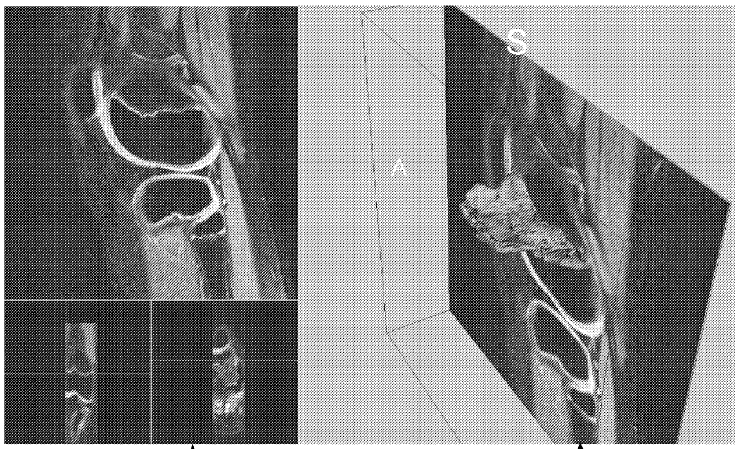
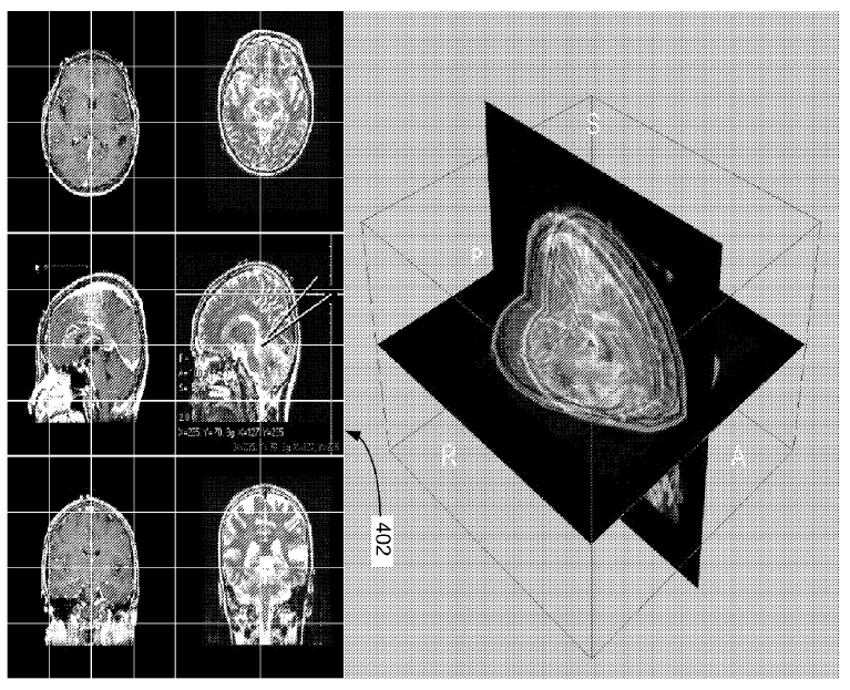
| D. Gering. System and Method for Flattened Anatomy for Interactive Segmentation and Measurement. US Patent #US7643662, General Electric Co., August 2006. |
| ABSTRACT: Systems and methods are provided for accessing three dimensional representation of an anatomical surface and flattening the anatomical surface so as to produce a two dimensional representation of an anatomical surface. The two dimensional surface can be augmented with computed properties such as thickness, curvature, thickness and curvature, or user defined properties. The rendered two dimensional representation of an anatomical surface can be interacted by user so as to deriving quantitative measurements such as diameter, area, volume, and number of voxels. |
|
| 2.3M |
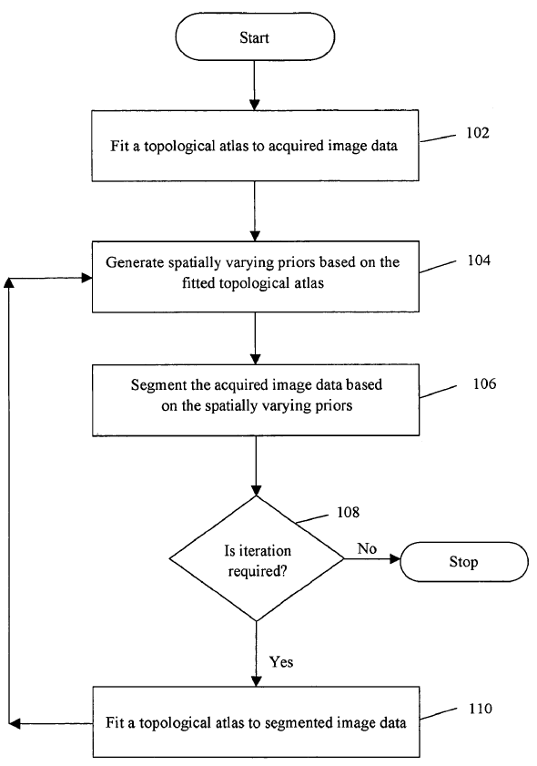
| D. Gering. Method and System for Segmenting Image Data. US Patent #US7623709, General Electric Co., September 2005. |
| ABSTRACT: Methods and system for segmenting image data are provided. The method includes fitting a topological atlas to acquired image data and generating spatially varying priors based on the fitted topological atlas. The method further includes segmenting the acquired image data based on the spatially varying priors. |
|
| 695K |
| D. Gering, G. Avinash, S. Gupta. Computer-Aided Detection System Utilizing Temporal Analysis as a Precursor to Spatial Analysis. US Patent #US7620227, General Electric Co., December 2005. |
| ABSTRACT: A computer-aided detection system for evaluating tissue based on a series of timed images acquired both before and after a contrast agent is administered performs a temporal analysis of the tissue and then a spatial analysis in which the tissue is categorized. After the temporal and spatial analysis is performed, the results can be displayed. The displayed results can include both tissue characterization results, underlying curves used to determine the characterization, and confidence data regarding how good the characterization is expected to be. The confidence data can be provided, for example, by using variations in color schemes, displaying numerical confidence levels, or providing graphical features such as piecewise linear models. |
|
| 827K |
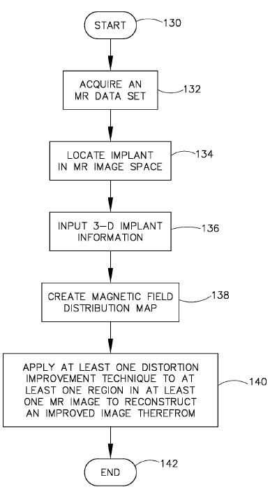
| K. Koch, R. Hinks, D. Gering. Method and Apparatus for Correcting Distortion in MR Images Caused by Metallic Implants. US Patent #US7535227, General Electric Co., October 2007. |
| ABSTRACT: A technique for reconstructing a corrected MR image from MR images distorted by foreign object induced magnetic fields includes locating a foreign object in a subject and defining a localized area of a field of view about the foreign object where a magnetic field distortion adversely affects a first magnetic distortion correction technique. The first magnetic distortion correction technique is applied to the field of view other than in the localized area. A second magnetic distortion correction technique is applied to the localized area and the results of the application of the first and second magnetic distortion correction techniques are combined. An image is reconstructed based on the results of the application of the first and second magnetic distortion correction techniques. |
|
| 1.0M |
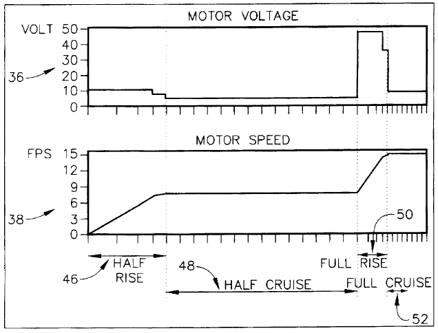
| D. Gering. System and Method of Motor Control. US Patent #5761376, General Electric Co., May 1997. |
| ABSTRACT: A method and system are provided for controlling a motor to rapidly accelerate and synchronize with an external synchronization pulse. First, the motor is accelerated to only half of the desired speed. At this point, the phase error with the sync pulse is calculated, and the motor continues "cruising" at half speed until the phase discrepancy diminishes. Then the motor rises to the full desired speed at the optimal time such that it is in phase with the sync pulse just as it reaches full speed. The acceleration is performed at a constant, yet slow enough, rate to receive at least three speed feedbacks. Then the instantaneous speed can be accurately measured and the next (and final) speed command during acceleration is interpolated between the ideal command for rising and the ideal command for cruising at the desired speed. The amount of phase discrepancy that diminishes while rising, as well as the ideal motor commands for cruising and rising, at both half and full speeds are adaptively learned and stored for the next run. |
|
| 580K |
Links