Research Projects of Samson Timoner I've been involved in many research projects -- and I have a lot of unpublished work. Let me begin by listing a few of the more recent projects. This was last updated in 2003:

Non-rigid Registration

Fast non-rigid registration of medical images is very difficult because there can easily be 8 million voxels in an image. Many algorithms require 3 to 4 hours to complete. We created a non-rigid registration that completes in 5 minutes, by changing the dominant computational complexity of the process. The computational complexity is dominated by the number of nodes in a tetrahedral mesh used to represent the displacement field, which using adaptive methods I created can be around 10 thousand nodes. The problem changes from order 8 million to order 10 thousand, and the registration problem can be solved in 5 minutes rather than 3 or 4 hours.
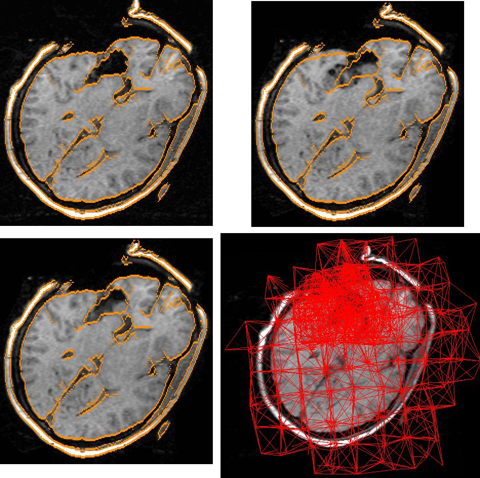
Top Left: Brain image 3 hours into surgery, with edges hilighted. Top Right: Brain image 4 hours into surgery, with the edges from the Top Left. Note that most of the disagreement is near the incision. Bottom Left: the same edges shown overlaying the deformed brain image -- the motion of the brain was accurately captured. 
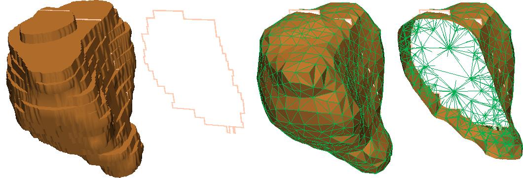
Bottom right: projection of the adaptive tetrahedral mesh. Note that the mesh is much more dense near the incision.

Segmentation

Intensity-based segmentation methods are very flexible, allowing complicated boundaries to be captured. However, they perform poorly in structures where there is a small intensity difference between the inside and outside of the structure. Deformable models perform well even in regions of poor intensity differences, but have difficulties capturing structures with widely differing structures. We created a way to merge the two methods. (Collaboration with Killian Pohl) Below you see a segmentation of the brain which focused on separation of white and grey matter, as well as capturing the right and left thalamus (magenta and violet), whose boundaries are very difficult to see

Classification

We created methods to separate normals and schizophrenic subjects based on the shape of sub-cortical structures. Our methods based on tetrahedral representations were shown to be significantly more effective at separating subjects than competing methods. Below is a map of how to take an Amygdala-Hippocampus complex from a diseased subject and make it look more like a complex from a normal subject. Red indicates an outward change, blue an inward change, and green no change.

Shape Matching

I created a fast algorithm to non-rigidly register shapes using tetrahedral representations. The algorithm treats objects like pieces of rubber and tried to minimize the stress-strain energy while deforming one shape into another. The results of this algorithm are useful for making deformable models for segmentation and for classification. Below are show left thalami where dots of the same color on each thalamus were found to correspond.

Tetrahedral Meshing

Tetrahedra are useful for describing 3D volumetric objects because tetrahedra can accurately describe smooth surfaces, and because a small number of tetrahedra can be used to describe the interior of an object. I wrote an algorithm to automatically fill medical objects with meshes of tetrahedra. Shown below is a prostate. From Left to Right: the surface of a voxel representation, a thin slice through it, then the surface of a tetrahedral representation and a thin slice through it.


Here are a few of the less recent research projects:
Samson Timoner: samsonaimitedu