
|
|
William M. Wells III
(aka Sandy Wells)
Professor of Radiology
Department of Radiology
Harvard Medical School and
Brigham and Women's Hospital
Member of the Affiliated Faculty of the Harvard-MIT Division of Health Sciences and
Technology
Research Scientist, MIT CSAIL
My BWH web page
|
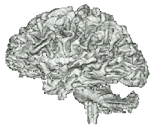
White Matter Surface
|
I am a researcher in medical image analysis with the
Surgical Planning Laboratory
, a unit of the MRI division of the
Radiology
Department
of
Brigham and Women's Hospital,
Harvard Medical School.
I maintain an active collaboration with
the
MIT Computer Science and Artificial Intelligence Laboratory,
where I have worked with a talented group of graduate students.
I am also affiliated with the
Harvard-MIT Division of Health Sciences and
Technology
, and periodically teach the medical image processing part of
HST882 / 6.555: Biomedical Signal and Image Processing
.
Modern medical images contain vast amounts of anatomical information.
Much of this information is accessible to diagnostic radiologists, in
part because people (in contrast to computers) are very good at image
interpretation. The anatomical information latent in such images is
also valuable for disease and neuroscience research, as well as for
drug trials. The quantitative analysis of medical images by computer,
however, remains challenging.
Among the most basic capabilities of medical image analysis are
segmentation
, the process of assigning labels to structures in images,
and
registration , the process of placing different images into
anatomical agreement.
My work has focused primarily on the analysis of structural
and functional MRI, including
segmentation and
registration of MRI, with some
emphasis on applications in image-guided surgery.
The figure on the upper right illustrates the white matter surface of a brain
that was segmented from MRI using Adaptive
Segmentation of MRI (the "EM Segmenter") .
My research in medical image registration concerns the use of
Mutual Information as a criterion for image
fusion. This approach has become the de-facto standard for multi-modality
problems.
Implementations of this method are available
in
3D Slicer,
our open-source platform for medical image analysis,
and in
ITK,
an NIH sponsored segmentation and registration library.
In addition to morphological analysis, I am also interested in
univariate and multivariate analysis of functional MRI.
I recently organized the 2013 meeting of Information Processing In Medical Imaging
(IPMI 2013)
, held near Monterey California June 29 - July 3.
Publications
My publications, with citation info, via Goole Scholar
My publications via the SPL Publications Database (some with pdf)
Contact info:

William Wells
Department of Radiology
Brigham and Women's Hospital
75 Francis St.
Boston, MA
02115
Directions to my office at Brigham and Womens Hosptial.
MIT office: 32-D462