The medical story of my hospitalization at
Mass General Hospital for 18 days in October 2009,
As I write this, I am nine days beyond open-heart surgery at MGH. In
this entry, I will describe my medical course. In future
entries, I plan to reflect on my experiences and to draw some
lessons learned from seeing the health care system from a patient’s
viewpoint.
Wednesday, October 14:
During a trip to Washington DC October 8-12, I experienced some
shortness of breath and tightness in my chest. Having had no history
of heart disease, I assumed this might be due to an upper
respiratory infection that others around me had experienced.
My symptoms abated to the point where I returned to normal work on
the 13th, but went to the MIT Medical Department on the 14th because
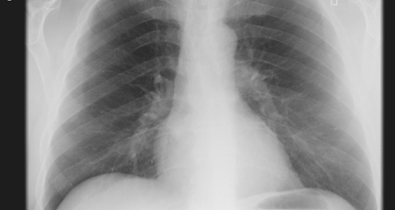
I still did not feel my usual self. After a completely normal
ECG and chest x-ray, Dr. David Shine at MIT listened to my history
and decided that despite the normal findings, the story sounded like
a new, hence by definition unstable, angina. He called an ambulance
to take me across the river to the MGH emergency department (ED).
I thought of my subsequent experiences as a kind of “Chutes and
Ladders” game, where successive tests led me up and, mostly, down.
After being wheeled in on a gurney, the ED put an ID band on my
wrist, gathered my insurance information, and moved me to a more
compact wheelchair for the obligatory four-hour wait until I could
be triaged, and then another two before my first lab test. I
later found out that that first test, a rapid test for troponin, was
normal (a ladder), but they kept me in the ED observation unit
overnight, where a more sensitive version of the test later in the
middle of the night revealed a mildly elevated troponin level of
0.09 (a chute), showing that some heart cells were leaking this
enzyme, probably in response to an inadequate supply of oxygenated
blood. This convinced them to admit me to a cardiac step-down
unit for further testing, cardiac monitoring, hourly vital signs,
and more blood draws seemingly every hour. Over the next day,
the troponin levels declined to 0.06, 0.06, and 0.04, at which point
they stopped measuring. One of the nurses also thought she heard a
pleural rub, which led to a null d-dimer test for pulmonary
embolism, but subsequent examination by others attributed it to
normal breathing sounds. In any case, I was told that a pericardial
rub would have been of much greater interest, but fortunately was
absent.
Thursday and Friday, October 15 and 16:
My first high-tech test was an ultrasound echocardiogram, performed
Thursday morning. It showed no abnormalities, and an ejection
fraction of 67%, which is even slightly above normal—a ladder. This
did, however, leave unexplained my symptoms and labs, so I was
scheduled for the next morning for a pharmacologic stress test.
I also met three cardiologists who had a wonderful guiding role in
my care. Dr. Shawn Gregory was my attending, Dr. David
Dudzinski, an HST MD grad, is his fellow and helped interpret what
was going on with my case above and beyond the call of duty.
Finally, my former student Zak Kohane, MD/PhD, introduced me to Dr.
Stanley Shaw, an HST MD/PhD, whom he knew from research connections,
but who is also an attending at my ward parts of the year.
Stanley has been an enormous help throughout the process, providing
me with information about my case not normally made available to
patients, inside “dope” on choosing practitioners, and simply being
available constantly to check on me, answer questions, talk through
options, etc. Finally, my old friend and colleague, Dr. Stephen
Pauker, offered wise advice while mostly traveling throughout this
period. Zak, Stanley and Steve included me in a spirited email
discussion that was just an ideal version of the kinds of
information and decision making that I have always believed patients
should be able to have with their providers.
The more common stress test uses exercise on a treadmill to put
stress on the heart, and looks for ECG changes. Because of my
recent history, they used what is called a pharmacologic stress test
instead. In this, I was first injected with a radioactive gamma
source, given time to allow that to perfuse, and then imaged by a
gamma ray camera, in the manner of a PET scan. Then, the same
procedure is repeated after I was also injected with a strong
vasodilator. The theory is that healthy vessels would dilate
and allow greater perfusion with the radioactive isotope, whereas
obstructed vessels cannot do this, so they would show relatively
poorer perfusion compared to the initial test. In fact, the
test showed “There is a small to moderate sized area of mild mid and
distal anterior, anteroseptal, and apical ischemia.” The rest of the
report was encouraging about it all being reversible, but those
areas sounded to me like they roughly correspond to the area of
heart muscle fed by the LAD (left anterior descending) artery. So my
expectation at this point was that I might wind up with a stent in
that vessel, and then deal with the downstream consequences of
Plavix and the risks of re-stenosis.
The next step at this point was a cardiac cathererization, but it
was too late to schedule it for Friday, which condemned me to a
weekend at “Spa MGH” with nothing to do but worry.
Monday, October 19:
The morning began early, with my transport to the cath lab, where I
got some mild anesthesia, was put on a high-tech table built around
a fluoroscope, was given a local anesthetic in my right groin, and
almost before I knew it, Dr. I. K. Jang had threaded a catheter into
my femoral artery and gotten a tube through the aortic arch and into
the starting points of the coronary arteries. Because of the
geometry of the table, I was unable to see the procedure, but within
an hour it was done, and the result was not what I had hoped for:
four-vessel disease, necessitating a coronary artery bypass graft,
or CABG. Clearly, another chute!
The following week provided less excitement, because open questions
had been resolved. The main question was how to schedule surgery,
and with which surgeon. I had a few recommendations for the
most famous MGH cardiac surgeons from my brother-in-law, Robert, who
had done a lot of work with them while he was a VP at Medtronic.
However, finding a surgeon is difficult, and these top folks are
mostly busy with far more complex surgeries such as cardiac
transplants, and would not be available to do my CABG for at least
several weeks. My local team recommended Dr. Jennifer Walker,
known for being thoughtful, fast, and having good outcomes.
She, too, was busy, but there was some chance we could get on her
dance card for Friday, though as it turned out the surgery was the
following Monday.
I met Dr. Walker on Friday, to discuss the surgery. She is a very
well-dressed, supremely self-confident woman, who I would guess is
in her 40’s. Of course, I think surgeons must be self-confident by
definition, but she also exudes competence. Dianne liked her
sense of style, thinking that she must do a neat job of surgery as
well. Dr. Walker explained that she planned to use my left internal
mammary artery to graft the LAD and pieces of the saphenous vein
from one of my legs to graft the three other blockages. Amazingly,
the body seems to have a great deal of redundancy, so it’s possible
to re-cycle arteries and veins without causing major problems. The
trickiest part of the procedure would be grafting the pieces of vein
into the aorta and then the one artery and veins to their
destinations. The plan was for surgery to last four hours “soup to
nuts”, and I heard that the “on-pump” time, i.e., with my heart
stopped to permit the core part of the surgery, was to take only 40
minutes. I was curious why this would be such a small fraction
of the total surgical time, but in retrospect I must have
(optimistically) misheard the estimate. [From the surgical
report, it seems that the operation took an actual five hours, with
140 minutes “on-pump”.]
There is no way to make this surgery sound pleasant. The leg
vein is harvested laparoscopically, which leaves only three small
incisions—a much better deal than I hear this surgery used to
require. However, access to the heart is obtained by sawing
the sternum apart, spreading the ribs and cutting through the
pericardial sac. During the core of surgery, the pump tubes
are grafted from the right heart to the aorta to provide circulation
of oxygenated blood to the body, my heart was to be stopped, and my
body temperature chilled to reduce complications. At the end, all
this is to be undone. Scary!
Except for some more x-rays, an ultrasound to see if my carotids
needed to be cleaned out during surgery, and another to choose my
left saphenous vein as the one to harvest for grafting, it was
mostly a waiting game, going by very slowly.
Monday, October 26:
The day of surgery finally arrived. The evening before, I had
been shaved and painted in betadine, a disinfectant. Around 6am, I
was wheeled to the OR, where Dianne was able to visit me before the
start. I was transferred to the operating table, given some
mild anesthesia, had an arterial line placed in my radial artery,
and engaged with one of the surgical team about how to remove my
wedding ring, which I had last put on when I weighed a lot less, but
which would now threaten to cut off circulation in my finger as I
got fluid loaded during surgery. That was the last thing I
remembered, until some very vague memories late that evening
recovering in the ICU, where I recall having brief conversations
with Dianne and Stanley, but no content. I later heard that my
surgery had taken about five hours.
Recovery, Tuesday to Sunday, October 27 to November 1:
According to plan, I should only have spent about a day in the ICU,
but because my blood pressure was slow to stabilize, I was there for
three days. Most of it is, thankfully, a blur. I was on
narcotics and various other pain relievers, aware of both doctors
and a few visitors, and vaguely worried about my continued need for
levophed, remembering that at least for general ICU patients,
inability to wean from pressors is a bad prognostic sign.
However, by Thursday I was out of the ICU, in the surgical step-down
unit, gradually losing my chest tubes, radial line, jugular vein IV,
another giant IV in my left arm, temporary pacer wires, etc.
By Friday, I could luxuriate in the shower, though I felt weak as a
kitten, and after proving that I could walk several tours of the
ward and climb a flight of stairs, I was discharged home on Sunday
evening.
Home:
My main responsibilities since being home are to continue to expand
my lungs by using an incentive spirometer (easy), coughing
occasionally (quite painful), and walking around the house and
neighborhood. Tylenol three times a day seems to do well enough at
controlling my sternal pain, where the sawn-apart sternum is wired
together by a permanently attached stainless steel wire that I am
told will not set off metal detectors or prevent MRI images. It’s
definitely no fun, but I’ve been out a bit every day, managed to
vote in yesterday’s municipal elections, and have been well cared
for by Dianne.
Overall, I went from thinking of myself as rather healthy, swimming
laps, walking and doing all I needed to do in life. It’s a
shock to accept that my heart was actually in deep trouble, as a
succession of tests showed the problems to be serially more
intense. Despite this, I feel fortunate not to have had a
serious heart attack that might have left me disabled or dead. And
as Steve Pauker says, I have essentially a new cardiac
vasculature that should give me many years of good service if I take
care of it. Dianne promises to help me work on that!
I also feel fortunate in the emotional support I have gotten from my
family, students and colleagues. Plus, my co-teachers have taken the
responsibility for teaching from my shoulders, and my colleagues
have stepped in to take over my research projects during my
recovery. Back to Blog